Retinal Detachment
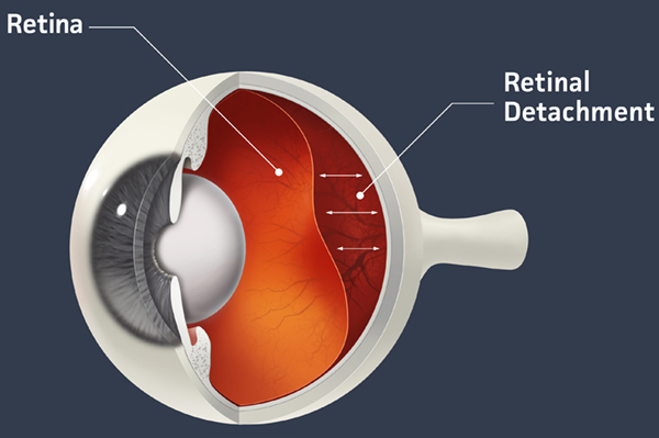
Retinal detachment occurs when the retina separates from the underlying layer of supporting tissue in the back of the eye. The primary cause of retinal detachment occurs from small holes or tears caused by factors such as trauma, prior intraocular surgery, or degenerations in the retina. Fluid from the center of the eye can enter these tears and holes and start to peel the retina off the back of the eye. Symptoms of retinal detachment include the development of floaters and flashes. Bleeding from the tear in the retina will cause floaters which can come in a variety of shapes. The pulling of the vitreous (the jelly inside the eye that shrinks as we age) will cause the flashes. As the retina detaches, images at the edge of your vision will become blurry. Patients experiencing any of the symptoms associated with retinal detachment, including floaters, flashes, and blurred or darkened vision, should contact an eye doctor for an examination.
There are several treatments that can be administered depending on the severity of the retinal detachment. If a tear or hole in the retina is found, it can generally be treated with laser surgery (retinopexy) or a freezing treatment (cryotherapy) to seal the tear by securing the retina to the back of the eye. If the retina has become fully detached, a silicone band, called a scleral buckle, will be attached to the eye forcing the retina back in place. The vitreous gel may also need to be removed in order to repair the eye; this can be achieved through a surgical procedure called a vitrectomy. If the retinal detachment has affected the vision in the center of the eye, it is likely that, even with surgery, this vision will not be restored. For this reason, it is important to seek immediate care if you believe you might be suffering from retinal detachment.